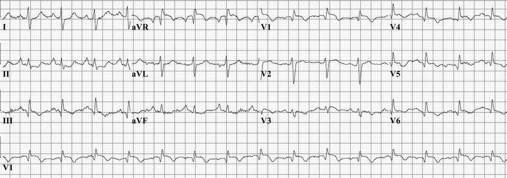
What is ‘S1Q3T3 sign’?
S1Q3T3 sign is Seen in ECG

R.W.Koster, CC BY-SA 3.0 https://creativecommons.org/licenses/by-sa/3.0, via Wikimedia Commons
- Image – points to note
- S wave present in lead -I
- Q wave and inverted T wave in lead III
S1Q3T3 sign reflects
[A] Right atrial strain
[B] Right ventricular strain
[C] Left ventricular strain
[D] Pulmonary artery strain
S1Q3T3 sign
S1Q3T3 sign
- Prominent S wave in lead I
- Q wave and inverted T wave in lead III
- This is a sign of acute cor pulmonale
- Suggests – acute pressure and volume overload of the right ventricle because of pulmonary hypertension
- Reflects right ventricular strain
S1Q3T3 findings on ECG and diagnosis of pulmonary embolism
S1Q3T3 findings on ECG – is present in 15% to 25% of patients ultimately diagnosed with pulmonary emboli
S1Q3T3 findings on ECG and acute cor pulmonale
Any cause of acute cor pulmonale can result in the S1Q3T3 findings on ECG,
- Pulmonary embolism
- Acute bronchospasms
- Pneumothorax
Pulmonary embolism – findings on ECG
S1Q3T3 findings on ECG
Other ECG findings noted during the acute phase of a PE include
- New right bundle branch block (complete or incomplete)
- Rightward shift of the QRS axis
- ST-segment elevation in V1 and aVR
- Generalized low amplitude QRS complexes
- Atrial premature contractions
- Sinus tachycardia
- Atrial fibrillation/flutter
- T wave inversions in leads V1-V4


